Projects in Progress
Constrained Disentanglement for CT Metal Artifact Reduction
The goal is to develop deep learning algorithms for CT metal artifact reduction in the context of proton therapy.
- Grant: NIH/NIBIB R01EB031102
- Multi-PIs: Ge Wang*, Bruno De Man (GE), Harald Paganetti (Harvard)
- Period: 09/01/2021 – 08/31/2025
- For more details, visit website
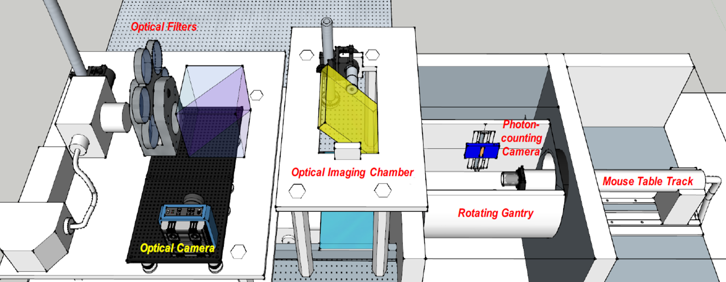
Photon-counting CT and Optical Molecular Tomography
The goal is to develop a preclinical x-ray and optical prototype for High-dimensional Optical Tomography (HOT) Guided-by Energy-resolved Micro-CT (GEM), visualize breast tumor heterogeneity, HER2 expression and dimerization, and therapeutic response in mice.
- Grant: NIH/NCI R01CA237267
- Multi-PIs: Ge Wang*, Xavier Intes, Barroso, Margarida (Albany Medical Center)
- Period: 09/01/2019 – 08/31/2024
- For more details, visit website
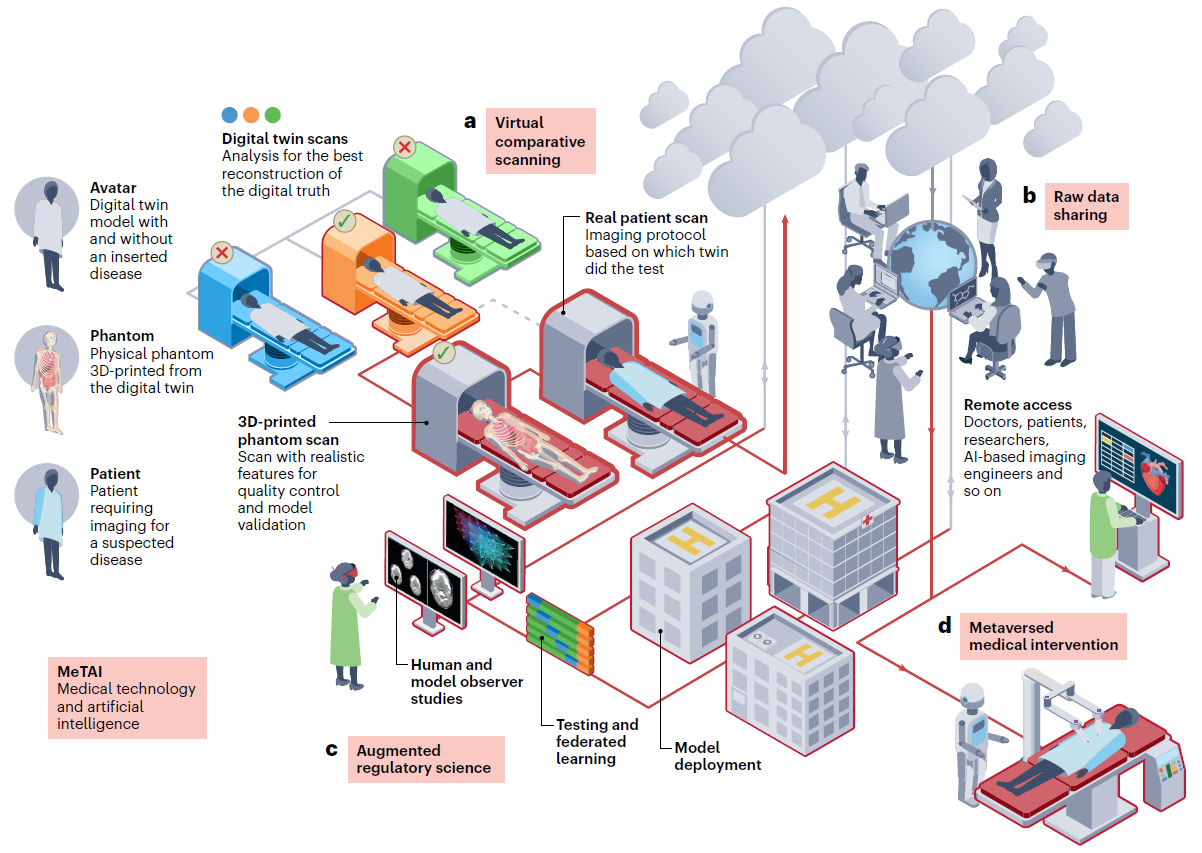
Adversarially Based Virtual CT Workflow for Evaluation of AI Imaging
The goal is to develop an FDA workflow evaluating, approving, and monitoring AI imaging software. This project would be a key element in the future healthcare metaverse.
- Grant: NIH/NIBIB R01EB032716
- Multi-PIs: Ge Wang*, Klaus Mueller (Stony Brook), Xun Jia (JHU), Rongping Zeng (FDA)
- Period: 04/01/2022 – 03/31/2026
- For more details, visit website
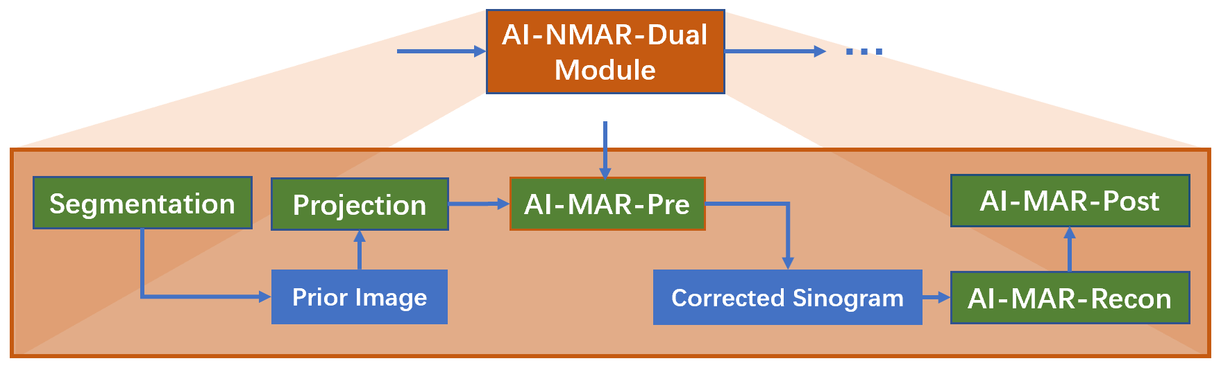
Cardiac CT Deblooming
The goal is to use deep learning to eliminate blooming artifacts in cardiac CT images without costly redesign of the CT hardware. This will be based on a dual-domain workflow from sinogram processing through image reconstruction to image analysis.

- Grant: NIH/NIBIB R01HL151561
- Multi-PIs: Bruno De Man* (GE), Ge Wang, James Min (Cleerly/Cornell)
- Period: 04/01/2020 – 06/30/2025
AI-based Cardiac CT
The goal is to develop an artificial intelligence (AI)-based approach to freeze the beating heart in CT images within a 60-millisecond time window (1/20 of a heart beat), eliminate the motion blurring in these images, and facilitate analysis of plaque buildup in these arteries.
- Grant: NIH/NIBIB R01EB032807
- PI: Hengyong Yu
- Subcontract PI: Ge Wang
- Period: 03/01/2023 – 02/28/2027
Unsupervised Deep Photon-Counting Computed Tomography Reconstruction for Human Extremity Imaging
The goal of this project is to develop an Unsupervised Deep Learning Approach (UDLA) for few-view and low-dose image reconstruction, with much higher spatial resolution and computational efficiency, without the requirement of ground-truth for training.
- Grant: NIH/NIBIB R01EB034737
- PI: Hengyong Yu
- Subcontract PI: Ge Wang
- Period: 07/01/2023 – 04/30/2027
SPECT with a Compton Camera for Thyroid Cancer Imaging
This goal is to design a high-efficiency and high-quality Compton camera based tomographic imaging system.
- Grant: NIH/NCI R21CA264772
- MPIs: Hengyong Yu*, Ge Wang
- Project schedule: 05/01/2021 – 08/30/2024
Radiomic Markers of Response and Recurrence for Cancer Patients
The goal is to develop and validate robust imaging features by standardizing image acquisition, improve automated tools for clinical trial use, and validate the predictive power of imaging features with external data.
- Grant: NIH/NCI R01CA233888
- Sub-PI: Wang
- Project schedule: 03/01/2019 – 08/31/2024
Deep Learning for Medical CT
The goal is to develop medical CT algorithms.
- Industrial grant: General Electric (USA)
- PI: Wang
- Project schedule: 01/01/2021– 12/31/2024
Investigation in Progress
Simultaneous CT-MRI Systems (Either High-end or Portable)
- Wang G, Zhang J, Gao H, Weir V, Yu HY, Cong WX, Xu XC, Shen HO, Bennett J, Furth M, Wang Y, Vannier MW: Towards Omni-Tomography – Grand Fusion of Multiple Modalities for Simultaneous Interior Tomography. PLoS ONE 7(6):e39700. doi:10.1371/journal.pone.0039700, 2012
- Wang G, Kalra M, Murugan V, Xi Y, Gjesteby L, Getzin M, Yang QS, Cong WX, Vannier MW: Simultaneous CT-MRI: Next chapter of multi-modality imaging. Med. Phys. 42:5879-5889, 2015
- Gjesteby L, Xi Y, Kalra M, Yang QS, Wang G: Hybrid imaging system for simultaneous spiral MR and X-ray (MRX) scans. IEEE Access, DOI:10.1109/ACCESS.2016.2637660, 2016
- Peng YT, Li MZ, Grandinetti J, Wang G, Jia X: Top-level Design and Simulated Performance of the First Portable CT-MR scanner. IEEE Access, doi:10.1109/ACCESS.2022.3208278, 2022
Auto-driving Vehicle for Affordable Tomo-Analytic Robots (AVATAR)
Given unprecedented progresses in the engineering field over the past decade or so, the development of AVATAR becomes feasible to integrate cutting-edge machine learning, auto-driving, medical imaging, robot, computer vision, healthcare metaverse, high-performance computing, and internet technologies, and will eventually change the landscape of the imaging world. This is particularly helpful for cancer screening, diagnosis, and follow-up in low-middle income countries (LMIC). See more details in the bilingual blog on AVATAR in Chinese (upper part) and English (lower part)